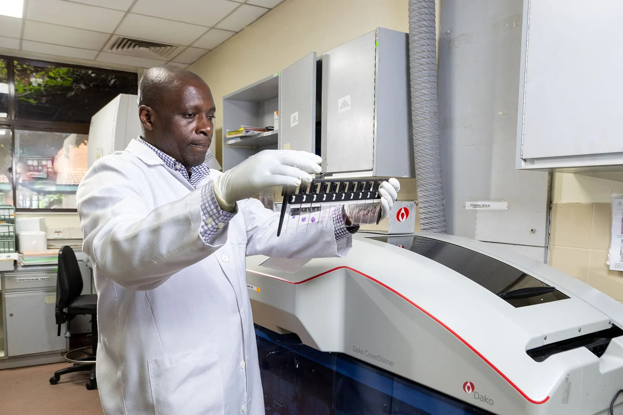
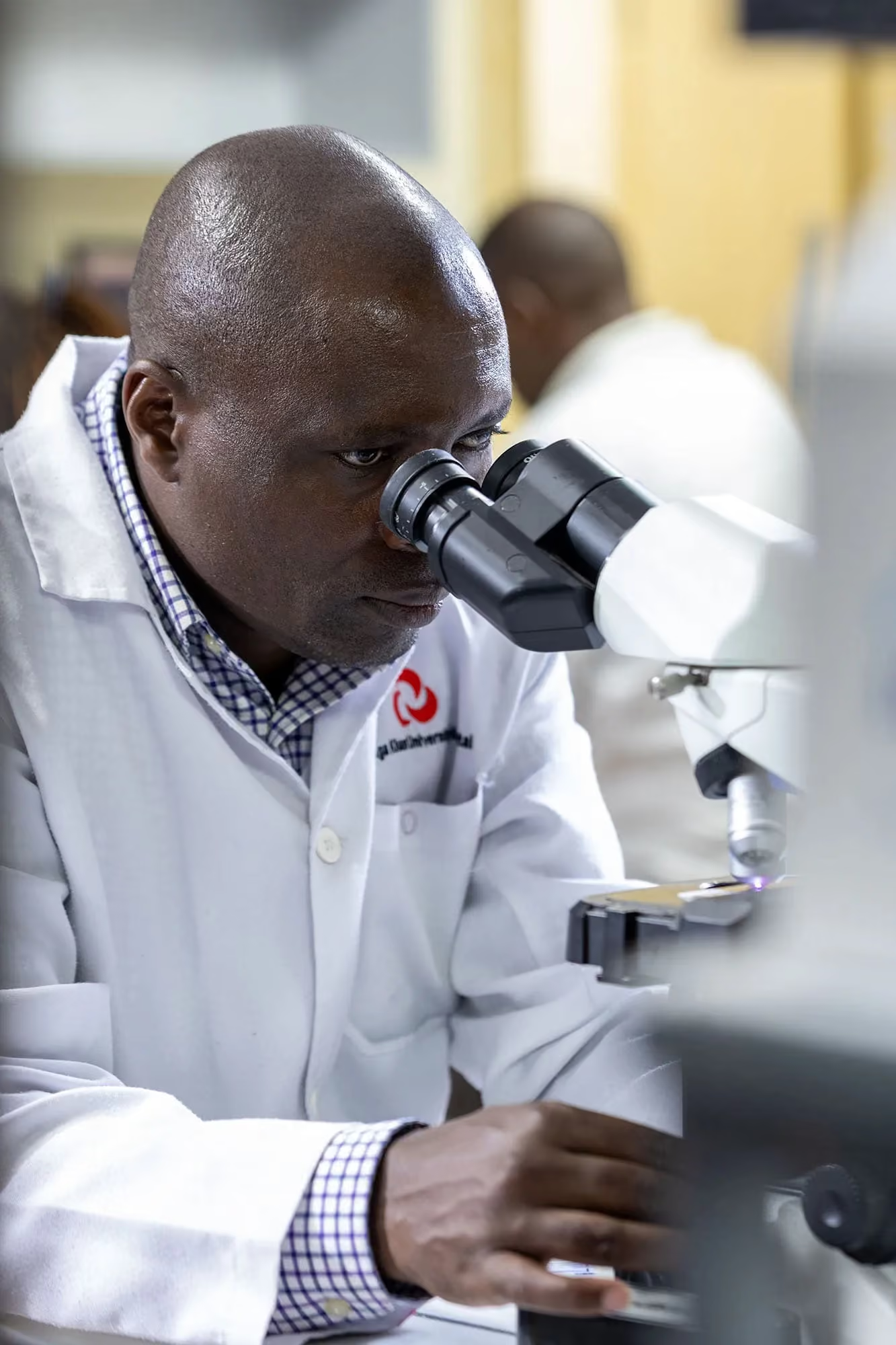
Shahin Sayed once thought her microscope would be a constant companion in the search for cancer. The Aga Khan University Hospital Nairobi (AKUHN) associate professor and chair of the Department of Pathology has often peered through its lens onto stained slides, searching for features that could lead to a patient’s cancer diagnosis.
Working from Nairobi, AKUHN Professor and Chair of the Department of Haematology-Oncology Mansoor Saleh are researching whether an AI tool could reenvision cancer diagnoses around the world and overcome pathologists’ dependence on microscopes and trained human eyes in diagnosing cancer.
The transition to AI is critical given that Africa has only 1/1000 of the number of trained pathologists compared to North America. Along with AKUHN Project Manager Alfred Mburu, they have partnered with University of Michigan Pathology Professor Ulysses Balis to test an AI technology he has developed over the last 18 years known as Vectorising Spatially Invariant Pattern Recognition (VIPR), which shows immense promise in identifying colon cancer cells on tissue specimens.
Akbar Waljee, who is the Leslie D. Yamada and Tachi Yamada M.D. Director, University of Michigan Center for Global Health Equity, facilitated the partnership and is a project co-principal investigator, along with Sayed, Balis, and Arvind Rao, associate professor of biostatistics and computation medicine and bioinformatics in the U-M School of Public Health. Saleh serves as principal investigator of the initiative.
Around 20% of Kenya’s colon cancer patients are under 40. However, the continent is also facing a severe shortage of pathologists to provide timely cancer diagnoses, with demand for their services far outpaced by patients’ needs. Sayed estimated there are around 120 pathologists in Kenya, a country with a population around 55 million. At the current rate of training, it would take almost 400 years to reach the number of pathologists per million people screened in the U.S.
While VIPR will not replace pathologists, whose expertise is required to guide the AI tool towards a diagnosis, it could lead to earlier cancer detection in a country where a majority of patients are diagnosed at later stages. Balis predicts pathologists will someday be able to create their own diagnostic algorithms for diseases beyond colon cancer.
“We’ve gotten excellent results in terms of the AI offering an accurate diagnosis. I absolutely think this will revolutionize the way we do diagnostics worldwide,” says Sayed.
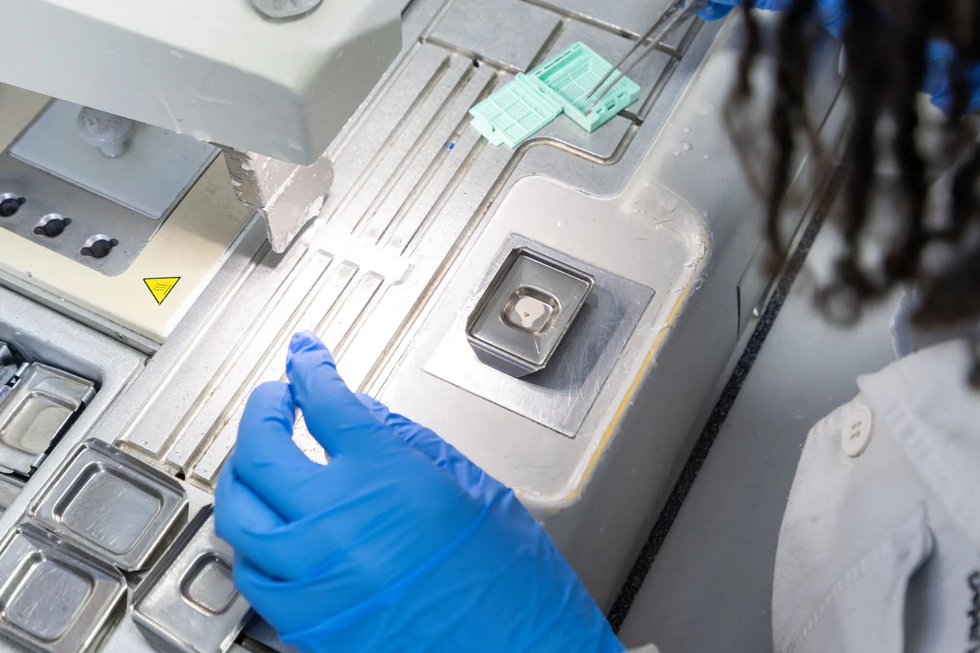
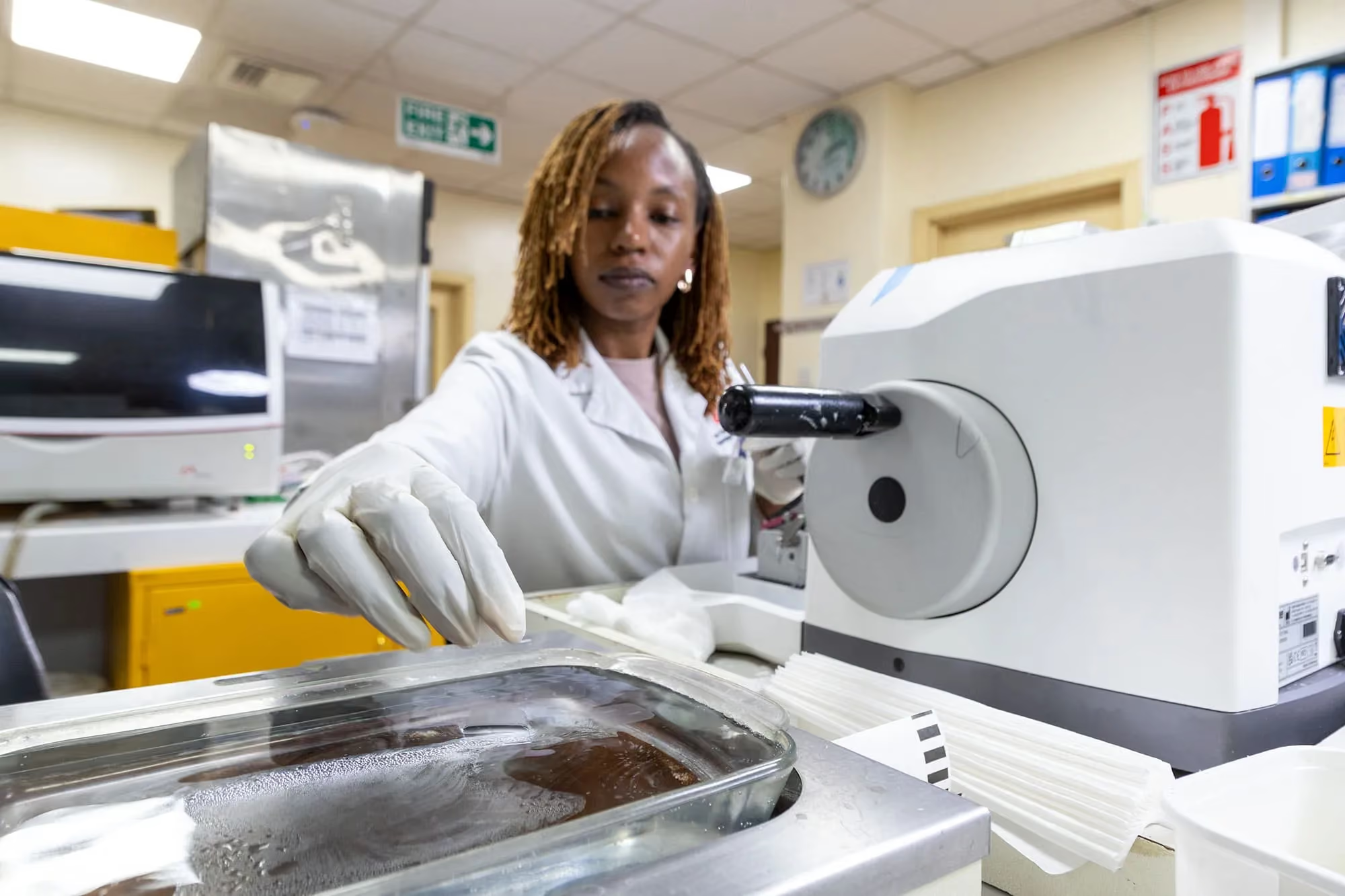
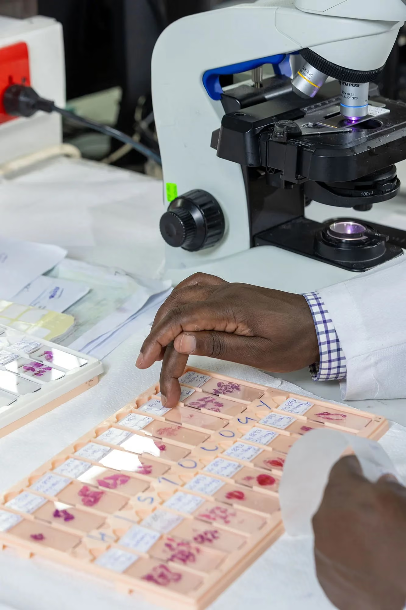
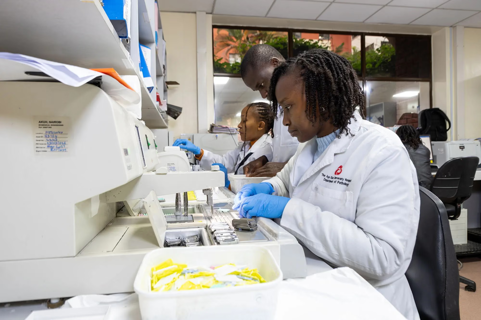
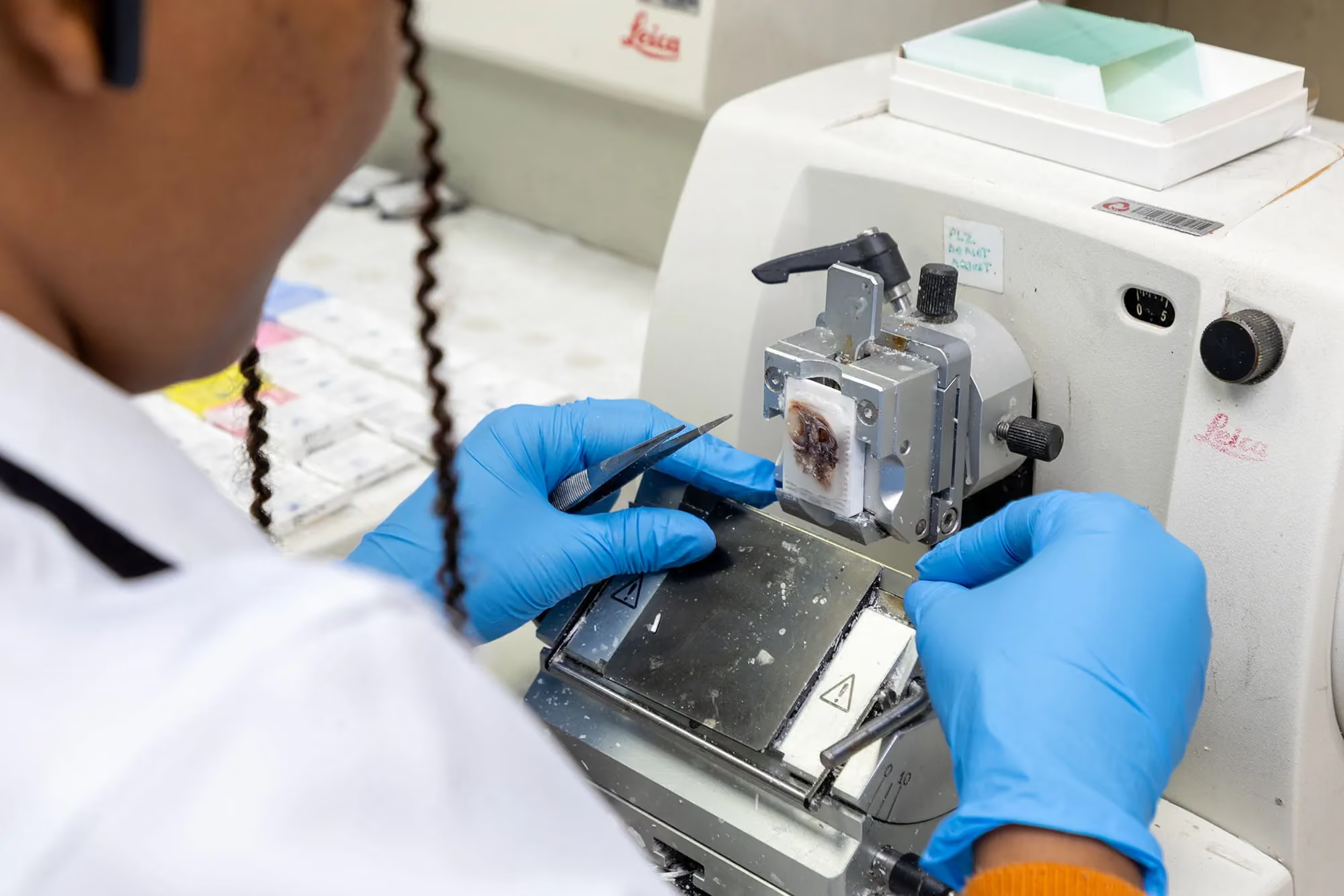
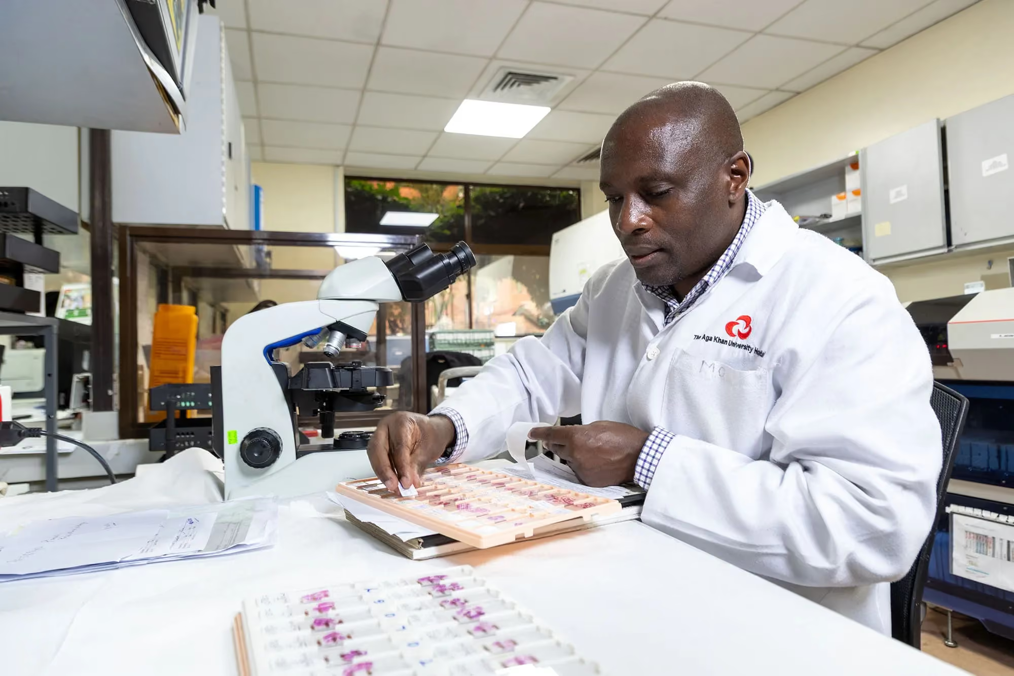
When pathologists manually search for cancer, they prepare thin slices of tissue collected during colonoscopies by placing them on small glass slides and staining the tissue with a purple dye called hematoxylin and an orange dye called eosin. They look through the microscope for features of colon cancer, which Sayed described as haphazardly arranged cells with large nuclei, among other features.Sayed and other pathologists have validated the VIPR-based AI tools with samples of local cases of colon cancer uploaded to a Microsoft Data Center in South Africa. The computational pipeline provided by Balis’ lab translates the digital images obtained from the glass slides into multi-dimensional information for the AI to analyze. VIPR assigns a biological meaning to each pixel of the image to determine what kind of tissue that pixel represents. When malignant cells are present, the AI can flag them within the image for pathologists to study and make a diagnosis.
The VIPR system differs from other AI tools. Rather than searching for overall patterns, it breaks images down into miniscule parts.
The technology can detect minute abnormalities in colon cells that might otherwise be a “needle in a haystack,” reducing the cognitive burden faced by pathologists with significant caseloads. The VIPR algorithm localizes areas of concern for pathologists to confirm, transforming the painstaking process of screening samples into an AI-enhanced review.
“You can imagine VIPR could ease my life as a pathologist, because it has the potential to make me much more efficient and accurate,” Sayed said.
The team’s next area of research may include additional AI and computational methods to assist clinicians with the difficult task of choosing the most effective treatments based on a tumor’s molecular features.“Aggressive cancers with high proliferation and metastatic potential that are unresponsive to therapy are correlated with specific immunohistochemical markers that indicate their aggressiveness,” Saleh explained. “Now, we want to know if these AI tools, using a simple stained slide, can also identify cancers that have poor prognoses.”
One of the greatest benefits of VIPR technology is what it does not require: infrastructure in every pathology clinic or additional work for an overburdened population of pathologists.
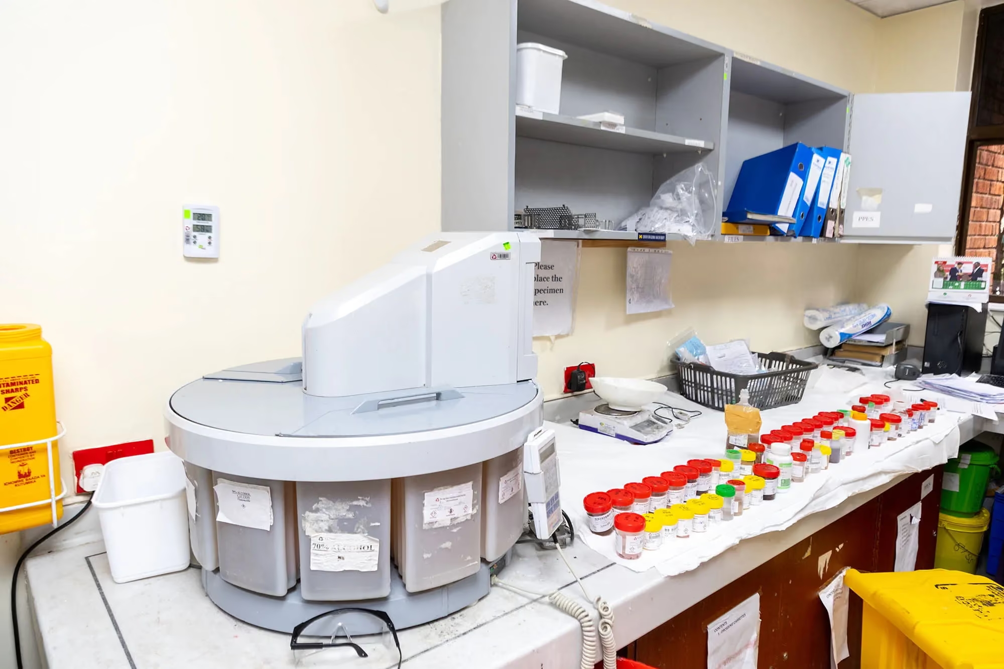
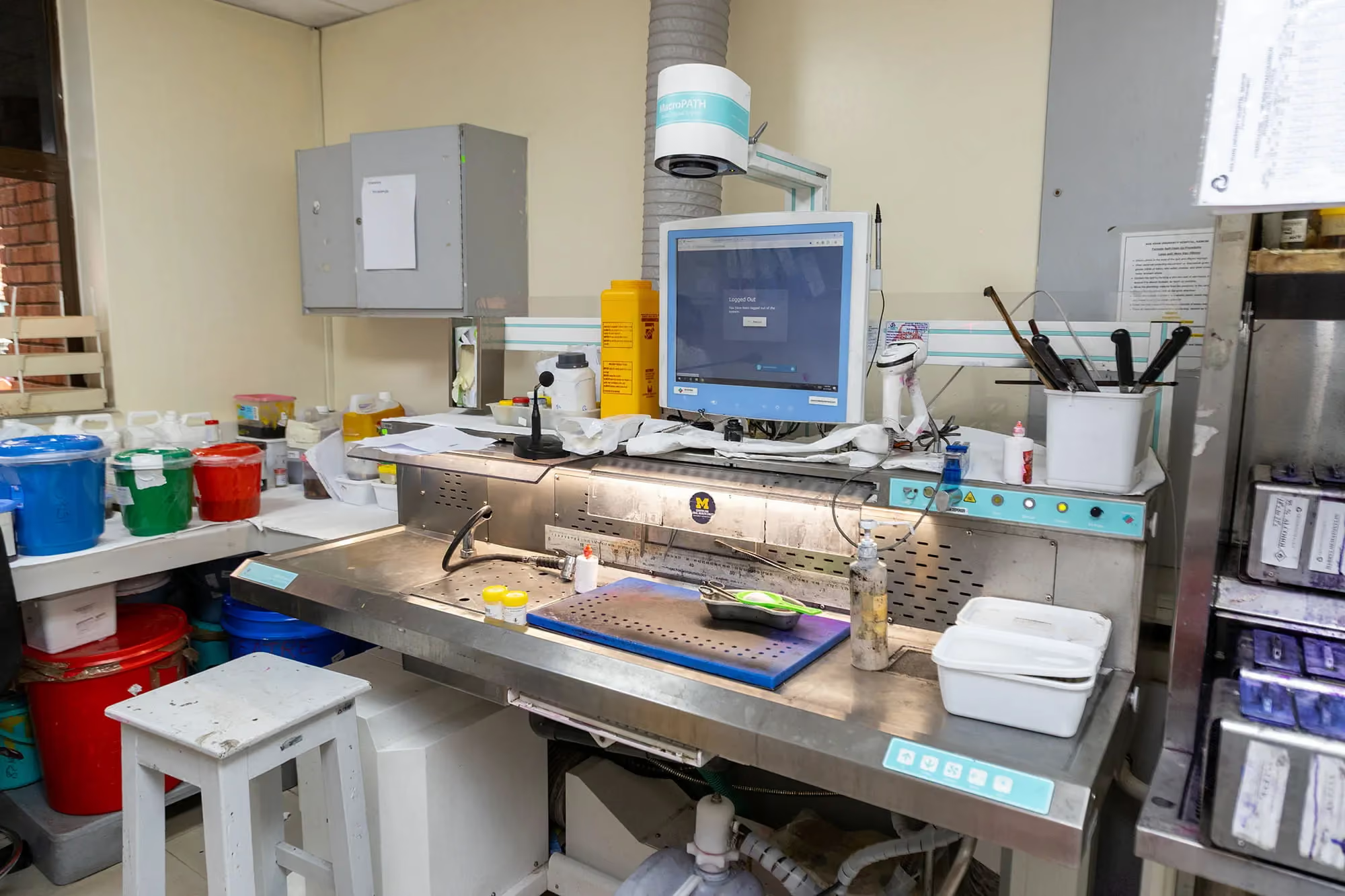
Currently, the AKUHN team digitizes and uploads slides using a digital microscope donated by U-M, which takes about one minute to upload each slide to the cloud. Saleh envisions investment in a centralized facility with increased cloud storage and faster scanners that can handle as many as 1,000 slides per hour to accelerate diagnostic tasks. Pathologists across the region could one day send locally scanned images of tissue samples to a central hub in Kenya for expedited analysis by AI tools, democratizing access for a larger segment of the population.
“We’ve been meeting with our government to discuss forming a cooperative to reduce equipment costs and maximize our impact,” said Saleh.
Pathologists don’t have to modify their workflows to use AI technologies like VIPR, which makes the tool a good match for low-resource areas, whether in Kenya or rural U.S. clinics.
They can collect tissue samples as usual, create the slides, and let the AI search for signs of cancer. Patients across the globe could benefit from expanded use of AI tools to deliver swift diagnoses, particularly those in regions with a shortage of pathologists.
Fortunately, training the AI is easy. Sayed has been supervising VIPR as it works through images of colon cancer cells. Pathologists can “handcraft” the cues AI uses for diagnosis by pointing out certain features, such abnormally shaped cell nuclei or tissue textures.
Many Kenyan patients aren't aware of colorectal cancer's early symptoms, which can be easily mistaken for schistosomiasis or other common gastrointestinal conditions, meaning some never seek screening in the first place. For those who do, access to colonoscopy remains limited, and without standardized record-sharing systems between providers, patients who relocate often fall through the cracks on follow-up care. By the time a diagnosis is made, many are living with advanced, treatment-resistant disease.
However, there is support for AI technology to improve patient care within the global pathology community. Sayed expected at least some pushback against moving from the microscope to the computer screen. But she has been pleasantly surprised to encounter a community of pathologists excited about the possibilities. AKU pathology residents are training with VIPR, and a faculty member will spend six months at U-M learning how to use the AI to create, train and deploy other AI algorithms and assist pathology labs in streamlining the process of using the tool.
The researchers are hoping computational pipelines like VIPR, as well as an expanding pool of additional solutions, may someday accurately assess how a patient’s cancer may progress and respond to treatment. Aga Khan University has follow-up data on 30 to 40 patients whose tissue samples are being used to train the AI. They’re also exploring the use of VIPR to develop and train an algorithm to identify breast cancer cells.
“It’s great to have this professional connection with U-M in service of patient care. I think the AI will really democratize healthcare spaces and make diagnostic tools more affordable and accessible.”